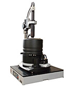
Scanning Probe Microscope Ntegra Aura
Product Features
- NTEGRA Aura allows measurements in low vacuum environment – 10-2 torr with the following advantages. Due to a cantilever Q-factor increase measurements are more sensitive, and they are possible without bad influence of surface adsorbate.
- Additional possibilities are provided by a temperature table with sample heating up to 300 °C and the accuracy of temperature maintenance 0.05 °C.
- Measurements in a controlled gas atmosphere are possible as well.
Scanning probe Microscopy |
STM/ AFM (contact + semi-contact + non-contact) / Lateral Force Microscopy / Phase Imaging/Force Modulation/ Adhesion Force Imaging/ Magnetic Force Microscopy/ Electrostatic Force Microscopy/ Scanning Capacitance Microscopy/ Kelvin Probe Microscopy/ Spreading Resistance Imaging/ Lithography: AFM (Force and Current), STM |
Specification |
Scan type |
Scanning by sample |
Scanning by probe* |
Sample size |
Up to 40 mm in diameter, to 15 mm in height |
Up to 100 mm in diameter, up to 15 mm in height |
Sample weight |
Up to 100 g |
Up to 300 g |
XY sample positiniong range |
5x5 mm |
Positioning resolution |
Readable resolution -5 um
Sensitivity -2 um |
Scan range |
100x100x10 um
3x3x2,6 um |
100x100x10 um
50x50x5 um |
Up to 150x150x15 um**(DualScan™mode) |
Non linearity, XY
(with closed loop sensors) |
≤ 0.1% |
≤ 0.15% |
Noise level, Z
(RMS in bandwidth 1000Hz) |
With sensors |
0.04 nm (typically),
≤ 0.06 nm |
0.06 nm (typically),
≤ 0.07 nm |
Without sensors |
0.03 nm |
0.05 nm |
Noise level, XY*** 0.2 nm
(RMS in bandwidth 200Hz) |
With sensors |
0.2 nm (typically),
≤ 03 nm (XY 100 um) |
0.1 nm (typically),
≤ 0.2 nm |
Without sensors |
0.02 nm (XY 100 um),
0.001 nm (XY 3 um) |
0.01 nm |
Closed-Loop Equivalent™ |
Noise level, XY
(RMS in bandwidth 200Hz) |
0.012 nm (XY 3 um) |
Noise level, Z
(RMS in bandwidth 1000 Hz) |
0.02 nm |
Zoom accuracy |
5% typically |
Optical viewing system |
Optical resolution |
1 um |
3 um |
Field of view |
4.5 - 0.4 mm |
2.0 - 0.4 mm |
Continuous zoom |
Available |
Available |
Temperature control |
Range |
From RT to +150°C |
Stability |
±0.005°C (typically), ≤ ±0.01°C |
Vacuum system |
Pressure |
10-2Torr |
Vibration isolation |
Active |
0.7-1000 Hz |
Passive |
Above 1 kHz |
* Scanning head can be configured to serve as a stand-alone device for specimens of unlimited sizes.
** Optionally can be expanded to 200x200x20 m.
*** Built-in capacitive sensors have extremely low noise and any area down to 50x50 nm can be scanned with closed-loop control.
 Printer Friendly Version Printer Friendly Version
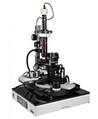
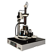
Scanning Probe Microscope Ntegra Maximus
Product Features
- NTEGRA Maximus basement is a changeable center unit with a motorized stage. Sample movement is possible within 50mm by X,Y and within 0-360 degrees under rotation.
- Automated measurements of many small samples are also possible with NTEGRA Maximus enabling high throughput screening.
- Macro language tool included in the base system provides highest possible flexibility in a research procedure selection and tuning.
- Scanning AFM head with capacitive sensors gives the system metrological properties.
Scanning Probe Microscopy |
AFM (contact + semi-contact + non-contact) / Lateral Force Microscopy / Phase Imaging/Force Modulation/ Adhesion Force Imaging/ Magnetic Force Microscopy/ Electrostatic Force Microscopy / Scanning Capacitance Microscopy/ Kelvin Probe Microscopy/ Spreading Resistance Imaging/ Lithography: AFM (Force and Current) |
Specification |
Sample size |
Up to 100 mm in diameter, up to 15 mm in height |
Sample weight |
Up to 1 kg |
XY sample positiniong range |
Linear movement range |
50 mm |
Positioning resolution |
2.5 um |
Rotary movement range |
360° |
Positioning resolution |
0.005° |
Scan range |
50x50x5 um |
Sample holder |
Vacuum chuck |
Non-linearity, XY
(with closed-loop sensors) |
≤ 0.15% |
Noise level, Z
(RMS in bandwidth 1000Hz) |
With sensors |
0.06 nm (typically), ≤ 0.07 nm |
Without sensors |
0.05 nm |
Noise level, XY*
(RMS in bandwidth 200Hz) |
With sensors |
0.1 nm (typically), ≤ 0.2 nm |
Without sensors |
0.01 nm |
Linear dimension estimation error
(with sensors) |
±0.5% |
Optical viewing system |
Optical resolution |
1 um/3 um |
Field of view |
4.5-0.4 mm |
Continuous zoom |
Available |
Vibration isolation |
Active |
0.7-1000 Hz |
Passive |
Above 1 kHz |
Built-in capacitive sensors have extremely low noise and any area down to 50x50 nm can be scanned with closed-loop control.
Printer Friendly Version
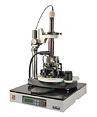
Scanning Probe Microscope Ntegra Prima
Product Features
- Equipped with a replaceable scanner with integrated capacitance sensors. A scan range of 100x100x12 um is available.
- XY nonlinearity: 0.05% of Peak-to-Peak by 2 value, after correction.
- Position repeatability in the X-Y plane is maintained with an accuracy of 10-20 nm in full scan range.
- The DualScan mode is performed with the use of the replaceable lower scanner (100x100x12 um) and another (100x100x10 um) upper scanner, which gives the total scan range of 200x200x22 um.
- The PNL controller and the mechanical parts of the PNL NTEGRA can operate in high-frequency modes, up to 5 MHz. This makes it possible to use the system both for AFAM applications and for operations with high-frequency cantilevers.
- This system can be used in research of high-resistance materials such as thin dielectric layers on semiconductors, DLC and piezo-films, conductive polymers etc.
Scanning probe Microscopy |
In air&liquid: AFM (contact + semi-contact + non-contact) / Lateral Force Microscopy / Phase Imaging/ Force Modulation/ Adhesion Force Imaging/ Lithography: AFM (Force) In air only: STM/ Magnetic Force Microscopy/ Electrostatic Force Microscopy/ Scanning Capacitance Microscopy/ Kelvin Probe Microscopy/ Spreading Resistance Imaging/ Lithography: AFM (Current), STM/ AFAM (optional) |
Specification |
Scan type |
Scanning by sample |
Scanning by probe* |
Sample size |
Up to 40 mm in diameter, to 15 mm in height |
Up to 100 mm in diameter, up to 15 mm in height |
Sample weight |
Up to 100 g |
Up to 300 g |
XY sample positiniong range |
5x5 mm |
Positioning resolution |
Readable resolution -5 um
Sensitivity -2 um |
Scan range |
100x100x10 um
3x3x2,6 um |
100x100x10 um
50x50x5 um |
Up to 200x200x20 um**(DualScan™mode) |
Non linearity, XY
(with closed loop sensors) |
≤ 0.1% |
≤ 0.15% |
Noise level, Z
(RMS in bandwidth 1000Hz) |
With sensors |
0.04 nm (typically),
≤ 0.06 nm |
0.06 nm (typically),
≤ 0.07 nm |
Without sensors |
0.03 nm |
0.05 nm |
Noise level, XY***
(RMS in bandwidth 200Hz) |
With sensors |
0.2 nm (typically),
≤ 03 nm (XY 100 um) |
0.1 nm (typically),
≤ 0.2 nm (XY 50 um) |
Without sensors |
0.02 nm (XY 100 um),
0.001 nm (XY 3 um) |
0.01 nm (XY 50 um) |
Linear dimension estimation error (with sensors) |
±0.5% |
±1.2% |
Optical viewing system |
Optical resolution |
1 um (0.4 um optional, NA 0.7)**** |
3 um |
Field of view |
4.5 - 0.4 mm |
2.0 - 0.4 mm |
Continuous zoom |
Available |
Available |
Vibration isolation |
Active |
0.7-1000 Hz |
Passive |
Above 1 kHz |
* Scanning head can be configured to serve as a stand-alone device for specimens of unlimited sizes.
** Optionally can be expanded to 200x200x20 m.
*** Built-in capacitive sensors have extremely low noise and any area down to 50x50 nm can be scanned with closed-loop control.
**** High Resolution Viewing system (HRV head) is optional and provides additional functionality making it possible to generate and detect tip-localized aperture less near-field effects.
Printer Friendly Version
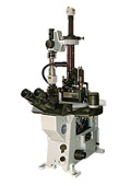
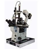
Scanning Probe Microscope Ntegra Solaris
Product Features
- New Shear Force head design offers:
-
100x100x10 um scan range
-
High scanning llinearity owing to integrated position sensors
-
Easy-to use Reflection mode realization
- The integrating of the inverted microscope objective into the central base becouse of high mechanical rigidity provides stability of the system making quality images and long-term experiments possible.
- Reflection and Transmission modes are available.
Scanning Near-Field Microscopy |
Shear Force Microscopy / SNOM reflection, transmission, luminescence (optional)/ any AFM modes are available optionally |
Specification |
Laser module |
Wavelength* |
441, 488, 514, 532, 633 nm |
Coupling unit |
X-Y-Z positioner, positioning accuracy 1 um |
V-groove fiber holder |
Coupling 40X objective |
Shear Force Imaging |
Sample size |
Up to 100 mm in diameter,
up to 15 mm in height |
XY sample positioning range |
5x5 mm |
Sample positioning accuracy |
Readable resolution-5 um
Sensitivity-2 um |
Closed-loop operation |
Capacitive sensors for 3 axes |
Scan range |
Scanning by sample |
100x100x25 um |
Scanning by probe |
100x100x7 um |
Non-linearity, XY |
Scanning by sample |
0.03 % (typically) |
Scanning by probe |
< 0.15 % |
Noise level, Z |
Scanning by sample |
< 0.2 nm (typically) |
Scanning by probe |
0.04 nm (typically), ≤ 0.06 nm |
Noise level, XY |
Scanning by sample |
< 0.5 nm (typically) |
Scanning by probe |
0.2 nm (typically), ≤ 0.3 nm |
Quartz tuning fork base frequency |
190 kHz |
Optical fiber diameter |
90 um (for 480-550 nm), 125 um (for 600-680 nm) |
Aperture diameter |
< 100 nm |
Channels for simultaneous registration |
Reflection |
Transmission/Fluorescence |
PMT detectors (for each channel) |
Spectral response |
185-850 nm |
Sensitivity at 420 nm |
3x10¹⁰ V/W |
Current-voltage conversion amplifier (built-in) |
1x10⁶V/A |
Frequency band width |
20kHz |
High voltage power supply |
Built-in |
Vibration isolation |
Active |
0.7-1000 Hz |
Passive |
Above 1 kHz |
* 488 nm laser is included as a default; other lasers can be supplied optionally.
Printer Friendly Version
Scanning Probe Microscope Ntegra Spectra
Product Features
- The universal PNL platform provides the possibility to integrate scanning confocal scheme in combination with regular AFM and spectrometer to detect Raman scattering spectrum. This spectra may then be interpreted into complex information concerning chemical composition of the object.
- Substantially extending optical microscope and AFM possibilities laser equipped system would be indispensable in environmental sciences, material sciences, living cell investigations.
Confocal Microscopy |
Optical module |
Inverted or upright microscope direct viewing system |
Housing with VIS optics (390-800nm) |
Polarizer in illuminator channel with Glan-Taylor prism 390-1000nm – manual |
Polarizer in detection channel with Glan-Taylor prism 390-1000nm – motorized |
1/2 wave plate, motorized – 3 position |
Beam splitter |
Evanescent excitation option (for TERS) |
Optical resolution |
XY |
200nm |
Z |
500nm |
Scanning module |
Sample weight |
Up to 1000g |
Scanning range |
100x100x25 um |
Closed-loop operation |
Capacitive sensors for 3 axes |
Non-linearity, XY |
0.03 % (typically) |
Noise level, Z |
< 0.2 nm (typically) |
Noise level, XY |
< 0.5 nm (typically) |
Pinhole |
Variable from 0 to 1,5 mm, step size 0,5 um |
Note that the sample for confocal microscopy can be either transparent or not and can be observed in air as well as in liquid environment.
Spectroscopy |
Spectrometer focal length |
520 mm |
Laser wavelength* |
441, 488, 514, 532, 633 nm |
Stray light rejection |
10-5 measured at 20 nm from 632 laser line |
Flat field |
28 mm x 10 mm |
Spectral resolution |
0.025 nm (1200 l/mm grating**) |
Ports |
1 input, 2 output |
Grating mounts |
4-position turret (3 gratings+mirror for "direct imaging" mode) |
Detectors |
CCD |
Spectral response 200–1000 nm, thermoelectric cooling down to –80°C, 95 % quantum efficiency at 500 nm |
Avalanche photodiode for photon counting*** |
Spectral response 400–1000 nm, dark counts = 25 counts/sec, supplied with PCI board with 1 GHz counting speed |
Printer Friendly Version
Scanning Probe Microscope Ntegra Therma
Product Features
- Special Thermohead™ provides extremely low thermal drift (less than 10 nm/°C) ensuring high stability of the tip-sample system. This allows long-term measurements to be done in pre-defined point on the specimen surface.
- Temperature control with sample temperature alteration is possible in the range of -30°C (Peltier element) to 200°C with the high temperature maintenance accuracy. It allows the observation of the structural changes on the specimen surface, such as crystallization, melting, growth processes, etc. with precise experiment conditions control.
- Different liquid cells are available: hermetic chemically stable liquid flow cell with a possibility of the temperature control at elevated temperatures; closed liquid flow temperature controlled cell for operating with Petri dishes; replaceable heater.
Scanning Probe Microscopy |
STM/ AFM (contact + semi-contact + non-contact) / Lateral Force Microscopy / Phase Imaging/Force Modulation/ Adhesion Force Imaging/ Magnetic Force Microscopy/ Electrostatic Force Microscopy/ Scanning Capacitance Microscopy/ Kelvin Probe Microscopy/ Spreading Resistance Imaging/ Lithography: AFM (Force and Current), STM |
Specification |
Scan type |
Scanning by sample |
Scanning by probe* |
Sample size |
Ambient environment |
Up to 40 mm in diameter,
to 15 mm in height |
Up to 100 mm in diameter, up to 15 mm in height |
Heating or cooling |
10x10x1.5 mm
15x12x1.5 mm |
Up to 15x17x1.5 mm |
XY sample positiniong range |
5x5 mm |
Positioning resolution |
Readable resolution -5 um
Sensitivity -2 um |
Temperature control |
Range |
From -30°C to +80°C/RT – +150 C |
From RT to 300°C |
Stability |
±0.005 (typically),
≤ ±0.01°C |
±0.01°C (typically),
≤ ±0.02°C |
Scan range |
-30°C - +80°C |
10x10x5 um |
- |
Ambient conditions/ RT - +150°C |
100x100x10um
3x3x2.6um |
50x50x5um |
RT - +300°C |
- |
50x50x5um |
DualScan™ mode |
Up to 150x150x15 um** (DualScan™ mode) |
Thermal drift***
(typically) |
XY |
15 nm/°C |
Z |
10 nm/°C |
Non linearity, XY
(with closed loop sensors) |
≤ 0.1% |
≤ 0.15% |
Noise level, Z
(RMS in bandwidth 1000Hz) |
With sensors |
0.04 nm(typically),
≤ 0.06 nm |
0.06 nm(typically),
≤ 0.07 nm |
Without sensors |
0.03 nm |
0.05 nm |
Noise level, XY****
(RMS in bandwidth 200Hz) |
With sensors |
0.2 nm (typically),
≤ 0.3 nm (XY 100 um)
0.025 nm(typically),
≤ 0.04 nm (XY 10 um) |
0.1 nm (typically),
≤ 0.2 nm |
Without sensors |
0.02 nm(XY 100 um),
0.002 nm(XY 10 um),
0.001 nm (XY 3 um) |
0.01 nm |
Linear dimension estimation error
(with sensors) |
±0.5% |
±1.2% |
Optical viewing system |
Optical resolution |
1 um/3 um |
3 um |
Field of view |
4.5-0.4 mm |
2.0-0.4 mm |
Continuous zoom |
Available |
Available |
Vibration isolation |
Active |
0.7-1000 Hz |
Passive |
Above 1 kHz |
Scanning head can be configured to serve as a stand-alone device for specimens of unlimited sizes.
** Optionally can be expanded to 200x200x20 m.
*** For temperature range –30°C – +80°C
**** Built-in capacitive sensors have extremely low noise and any area down to 50x50 nm can be scanned with closed-loop control.
Printer Friendly Version
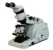
Scanning Probe Microscope Ntegra Tomo
Product Features
- Leica EM UC6 Ultramicrotome makes nanoslices of a sample and a freshly cut surface is then measured by AFM. Thus, many physical properties of a surface are studied and 3D-volume imaging is available after the reconstruction.
- After the NTEGRA Tomo operation sample slices stay available for TEM analysis
Scanning Probe Microscopy |
in-situ: AFM (contact + semi-contact + non-contact) / Lateral Force Microscopy / Phase Imaging/Force Modulation/ Adhesion Force Imaging/ Magnetic Force Microscopy/ Electrostatic Force Microscopy / Scanning Capacitance Microscopy/ Kelvin Probe Microscopy/ Spreading Resistance Imaging/ Lithography: AFM (Force and Current) |
Sample size |
Up to 8 mm in diameter,
up to 15 mm in height |
Sample weight |
Up to 10 g |
Scan range |
50x50x5 um |
Positioning resolution |
Readable resolution -5 um
Sensitivity-2 um |
Non-linearity, XY |
< 0.15% |
Noise level, Z
(RMS in bandwidth 1000 Hz) |
0.06 nm (typically), ≤ 0.07 nm |
Noise level, XY
(RMS in bandwidth 200 Hz) |
0.1 nm (typically), ≤ 0.2 nm |
Vibration isolation |
Dynamic |
Frequency range 0.7 – 1000 Hz |
Passive |
For frequencies above 1 kHz |
Ultratomy |
Self locking |
Yes |
Graduation |
±30° graduation |
Clearance angle adjustment |
-2° to 15° with 1°scale |
Knife holder |
For 6-12 mm knives |
Coarse knifemovements |
N-S |
10 mm stepping motor |
E-W |
25 mm stepping motor |
Cutting window |
0.2-15 mm adjustable |
Cutting speed |
0.05-100 mm/s wheel contr. |
Section thickness |
0-15000 nm wheel contr. |
FEED / SPEED storage |
5 |
Return speeds |
10, 30, 50 mm/s |
Step control |
0.1-15 m steps |
Section counter |
Yes |
Feed totalizer |
Yes |
Count down |
Yes |
Rocking mode |
Yes |
E-W measurement |
Yes |
Auto trim |
Yes |
Specimen advance indicator |
Yes |
Working distance |
110 mm |
Universal specimen holder |
2pcs. |
Flat specimen holder |
1p. |
Instrument table |
Dimensions |
Shock-absorbing elements |
Printer Friendly Version
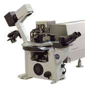
Scanning Probe Microscope Ntegra Vita
The integrating of the inverted microscope objective into the central base becouse of high mechanical rigidity provides stability of the system making quality images and long-term experiments possible. Inverted optical microscope stability is not so important any more.
Product Features
- Different liquid cells are available: hermetic chemically stable liquid flow cell with a possibility of the temperature control at elevated temperatures; closed liquid flow temperature controlled cell for operating with Petri dishes; replaceable heater.
- Hermetic liquid cell (MP4LCNTF)
- Flow-through possibility and heating (up to 60oC stability ~0,05°C)
- Control and changing of biochemical environment in solution during measurements
- Range of sample positioning 1x1 mm, positioning resolution 5 um.
- Chemically stable materials (stainless steel, fluoroplast) allow operation in aggressive solvents such as H2SO4, HCl.
- Quasi-hermetic liquid cell for Petri dishes (MP5LCNTF)
- Flow-through possibility and heating up to 50°C.
- Range of sample positioning 1x1 mm, positioning resolution 5 um.
- Removable heater for the separate cleaning. The heater and the temperature control sensor are located inside the fluoroplastic tube.
- Changeable probe holder with a glass prism and a silastic membrane
Scanning Probe Microscopy |
SPM methods |
in air & liquid |
AFM (contact + semi-contact + non-contact) / Lateral Force Microscopy / Adhesion Force Imaging/ Force Modulation/ Phase Imaging/ AFM Lithography (scratching)/ Force- Distance curves |
in air only |
STM/ Magnetic Force Microscopy/ Electrostatic Force Microscopy / Scanning Capacitance Microscopy/ Kelvin Probe Microscopy/ Spreading Resistance Imaging/ Lithography: AFM (Current), STM |
|
Scanning by sample |
Scanning by probe* |
Sample size |
in air |
40 mm in diameter,
15 mm in height |
100 mm in diameter,
15 mm in height |
in liquid |
Up to 14x14x2.5 mm |
Up to 15x15x3 mm |
XY sample positioning range |
in air |
5x5 mm, readable resolution -5 um
sensitivity -2 um |
in liquid |
1x1 mm, readable resolution -5 um
sensitivity-2 um |
Scan range |
100x100x10 um, 3x3x2.6 um
100x100x10 um, 50x50x5 um |
Up to 200x200x20 um** (DualScan™ mode) |
Non-linearity, XY
(with closed-loop sensors***) |
< 0.1% |
< 0.15% |
Noise level, Z
(RMS in bandwidth 1000 Hz) |
With sensors |
0.04 nm (typically),
≤ 0.06 nm |
0.06 nm (typically),
≤ 0.07 nm |
Without sensors |
0.03 nm |
0.05 nm |
Noise level, XY***
(RMS in bandwidth 200 Hz) |
With sensors |
0.2 nm (typically),
≤ 0.3 nm (XY 100 um) |
0.1 nm (typically),
≤ 0.2 nm (XY 50 um) |
Without sensors |
0.02 nm (XY 100 um)
0.001 nm (XY 3 um) |
0.01 nm (XY 50 um) |
Temperature control
(For operation in fluid environment) |
Range |
- |
from RT to 60°C |
Stability |
- |
≤ ±0.01°C |
*Scanning head can be configured to serve as a stand-alone device for specimens of unlimited sizes.
** 200 m scan range is possible with the unique DualScan™ mode when scanning by sample and scanning by probe can be done simultaneously.
*** Built-in capacitive sensors have extremely low noise and any area down to 50x50 nm can be scanned with closed-loop control.
Printer Friendly Version |